Multimodal imaging by elemental and molecular mass spectrometry
European Innovation Center – Interview with Prof. Uwe Karst, University of Münster (Germany)
Shimadzu’s European Innovation Center (EUIC) merges cutting-edge analytical technologies of Shimadzu with game-changing topics and expertise from leading scientists. This innovation-oriented cooperation focuses on creating new solutions for tomorrow.
The European Innovation Center applies a decentralized structure all over Europe to be in close local proximity to scientists and related markets.
With their leading-edge research expertise, highly-reputed scientists from well-known European universities cover the academic part of the EUIC. Their scientific focus areas include clinical applications, imaging technology, food and composites, with an emphasis on new methods, tools, techniques, diagnostics and solutions.
This issue of Shimadzu NEWS covers an interview with Professor Uwe Karst, University of Münster, Germany. With a focus on imaging technology within the EUIC, he gives insights on his current research projects.
To start, can you outline the research you conduct in general? What is currently state-of-the-art?
Our group specializes in the development and application of analytical methods and instrumentation to address complex analytical problems, originating mainly from the biomedical area.
One of our major research areas is speciation analysis. The need for speciation analysis is caused by the fact that the physiological properties (uptake, distribution, metabolism and excretion) of metal-containing compounds are strongly dependent on the individual metal species rather than on the heteroatom in general. This is easily proven using the example of hexavalent chromium, which is considered to be toxic and carcinogenic, while trivalent chromium is regarded as a possible essential trace metal species.
A second major research area of our group is chemical imaging – analysis of the distribution and spatially resolved quantification of low molecular weight analytes in biological matrices.

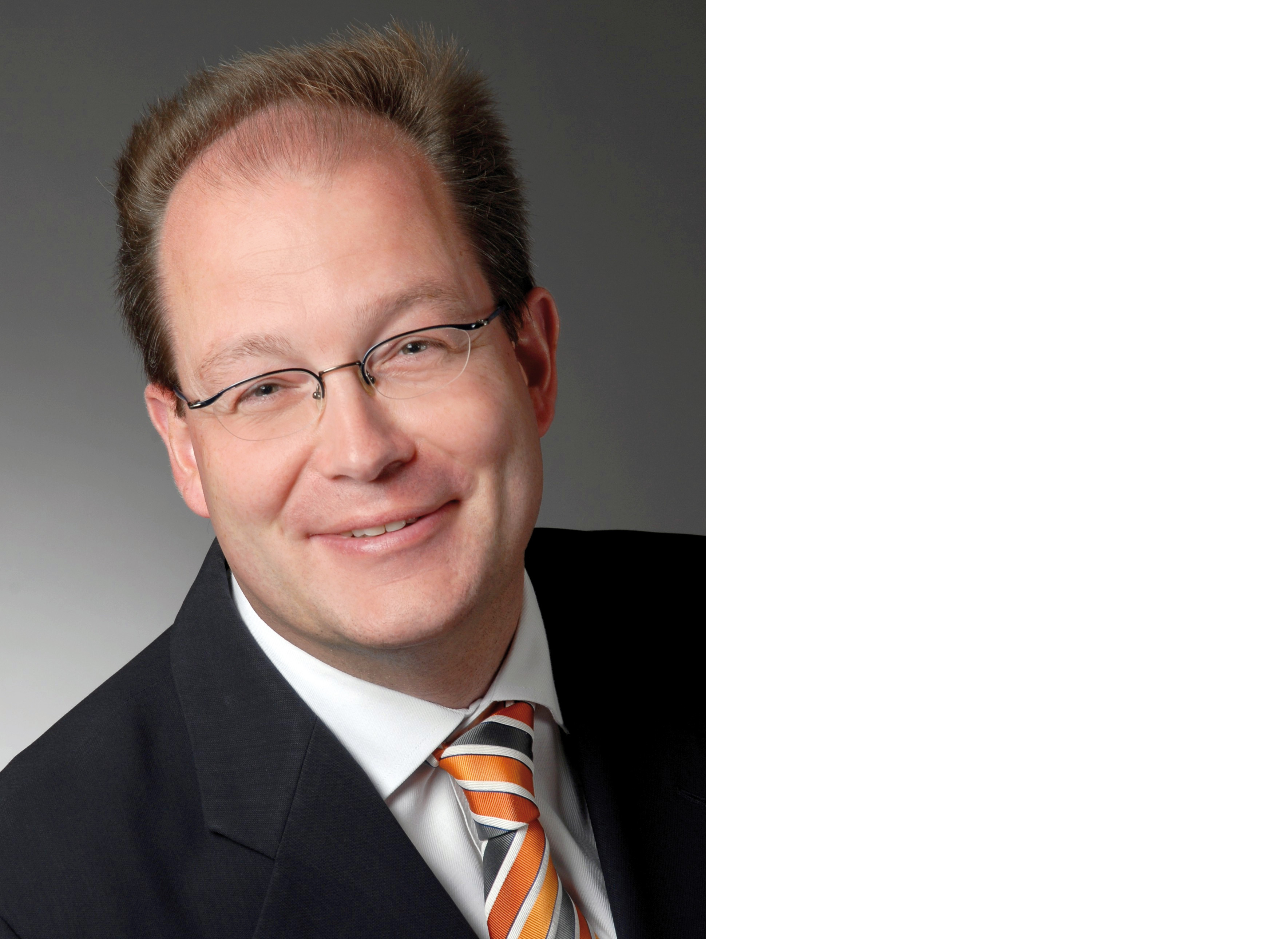
Uwe Karst holds the Chair of Analytical Chemistry at the University of Münster in Germany. After diploma and Ph.D. studies in Münster, which he finished in 1993, he joined the University of Colorado in Boulder as postdoctoral associate. He returned to Münster to obtain his habilitation and was appointed as Full Professor of Chemical Analysis at the University of Twente in the Netherlands from 2001 to 2005, after which he assumed his current position.
He is author of more than 250 publications in peer-review journals and 18 patents. Together with his research group, Prof. Karst has organized several international and national conferences including the International Symposium on Chromatography in 2008, the Metallomics Conference in 2011 and the European Winter Conference on Plasma Spectrochemistry in 2015.
How much are speciation analysis and chemical imaging linked to each other?
Speciation analysis and chemical imaging complement each other perfectly regarding the analysis of metal species in the organisms of humans, animals and plants.
Speciation analysis mostly uses liquid phase separations to separate the metal or metalloid species prior to identification by electrospray mass spectrometry (ESI-MS) and quantification by inductively coupled plasma-mass spectrometry (ICP-MS).
Chemical imaging, on the other hand, is carried out by matrix-assisted laser desorption/ionization-mass spectrometry (MALDI-MS) and related techniques to obtain distribution information about intact molecules, while distribution and quantification of the elements is accessible by laser ablation (LA) coupled to ICP-MS and micro-X-ray fluorescence (µXRF). As an example, investigation of side effects of Cisplatin-based tumor therapy requires speciation analysis to detect and quantify the individual platinum species formed in body fluids and tissues, while imaging by LA-ICP-MS provides quantitative distribution information on platinum in human or animal tissues.
Can you describe the research you are doing at the European Innovation Center with Shimadzu?
We have been collaborating with the EUIC on imaging for more than a year, and a major topic at this moment is further improvement of the combination of laser ablation and ICP-MS for chemical imaging purposes. As in other previous and current cooperations with various instrument manufacturers, we see our role not only in developing applications, but also in contributing to the further development of the actual and future instrument generations. This also includes suggestions for improved hardware, software for instrument control, data evaluation and integration of the instrument´s data with data of other imaging methods. Within our current project, the focus is directed on LA-ICP-MS and its combination with MALDI-MS and MALDI-MS/MS, as many of our current analytical challenges require the combined use of complementary imaging techniques.
Why are you interested in this research? What is the goal? Why is it important?
The analysis of low molecular weight compounds with physiological effects is currently (and probably always will be) an area of high complexity and individual analytical solutions. In contrast to Omics techniques, the potential for automation of procedures for high-throughput analysis is limited due to the strongly varying chemistry behind each analytical problem we are facing.
However, this chemical variability and the need to address challenges with a combination of technology and chemistry is exactly what I like in this field of research. For each question, there is a plethora of possible approaches to be checked, and interdisciplinary teams with colleagues from Medicine and Biology have to cooperate well to be successful. Even during my postdoctoral times, I would never have expected to be able to contribute to investigations on the side effects of platinum cancer chemotherapy, on the deposition of gadolinium from magnetic resonance imaging (MRI) contrast agents in the human brain or on fibrosis caused by certain types of nanoparticles in the lung.
Even more exciting, the combination of chemical and medical imaging opens up completely new routes for research. MRI, computed tomography (CT) or positron emission tomography (PET) are highly complementary regarding in vivo/in vitro situation, spatial resolution, limits of detection and capabilities for quantification.
How do Shimadzu instruments support your research?
As stated, combined imaging techniques are particular intriguing. Cooperation with manufacturers active in the fields of chemical imaging are therefore particularly attractive to us (and hopefully for the manufacturers as well). I just came back from a large Radiology congress which Shimadzu also attended, presenting equipment for medical imaging at their booth. While we have been using chromatography (LC, GC) and spectroscopy (UV/vis, fluorescence, AAS) instrumentation from Shimadzu for more than 20 years for research and education, our cooperation in the imaging field was established only three years ago, and is currently centered around LA-ICP-MS and complementary MALDI-MS work.
What are Shimadzu’s strengths compared to other vendors (not limited to the instruments)?
Cooperation is always based on trust and on personal relations, and a major reason for us to work with Shimadzu equipment at a larger scale during my habilitation phase, in which I had very limited equipment, was an excellent relation with the local sales agent. He was always helpful to a much larger extent than we could expect, and he was just an outstanding ambassador for the company.
Of course, well-performing and robust instrumentation helps as well, but the more complex the analytical challenges become, the more important is excellent communication and cooperation between individuals on both sides. Additionally, in our current situation, a broad spectrum of imaging instrumentation from any manufacturer is particularly attractive to us due to the complexity of our analytical problems and the increased chances to tackle the most difficult challenges.
Could you share any requests that you have with respect to analytical and measuring instrument vendors?
Let me start with a very general statement that is not addressed to any particular vendor: While the world of analytical challenges is converging more and more, even including a vanishing “wall” between “organic” and “inorganic” analysis, there are often business decisions of instrument vendors that lead to fragmentation of product lines and separation of business units that are hard to understand in the light of the increasing complexity of analytical challenges. Sometimes I wish that scientists were more involved in business decisions of instrument vendors, as this is not just a matter of rapid sales but also of long-term business relations and business development in a complex market situation. It may be naive to think that sometimes, wise long-term decisions should overrule rapidly profitable decisions, but it would accelerate technical progress so much …
Back down to earth again, my wish would be that manufacturers improve integration of their major product lines to a larger extent, which would be beneficial especially in our research areas of speciation analysis and chemical imaging. This is one of the major factors we are trying to contribute to in our cooperation with the Shimadzu European Innovation Center on imaging.
Take a look into the future: What will happen in the imaging field and how will the change influence the instruments/procedures in ten years?
In my opinion, this goes very much in line with my reply to your earlier question: I am aware that your major markets of today are not the research areas which we are currently working on and that your sales will go mostly to the mass markets in routine labs. However, we are facing an increasing degree of complexity, and being prepared to address this situation will become even more important in the future. While the medical imaging area will continue to expand due to its immediate and obvious benefit for the patient/customer, the chemical imaging area is harder to predict, as the immediate necessity for the paying customer is not as clear, thus hampering large investments in this field.
Regarding scientific content, there will be massive progress in chemical imaging, leading to strong optimism regarding future development. However, reaching the mass markets in routine labs (medical, environmental, food) will require a significant reduction in cost per analysis and for the need for highly qualified personnel. This can only be achieved by strong improvements in speed of analysis (more spots per second in imaging mode), improved hard- and software integration and possibly even strategic alliances between vendors of complementary instrumentation or between different divisions of one vendor.
Let me conclude with the statement that there is an increasing amount of light on the horizon, but that the full sunrise has to be earned by hard work and smart decisions in academia and industry. We will do our best to contribute!